
Medical Terminology Daily (MTD) is a blog sponsored by Clinical Anatomy Associates, Inc. as a service to the medical community. We post anatomical, medical or surgical terms, their meaning and usage, as well as biographical notes on anatomists, surgeons, and researchers through the ages. Be warned that some of the images used depict human anatomical specimens.
You are welcome to submit questions and suggestions using our "Contact Us" form. The information on this blog follows the terms on our "Privacy and Security Statement" and cannot be construed as medical guidance or instructions for treatment.
We have 418 guests online
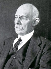
William S. Halsted, MD
(1852 – 1922)
American anatomist, teacher, and surgeon, William Stewart Halsted was born in New York City, USA to a wealthy family of English origin. His father was involved in charitable work and Governor and trustee to a city hospital. Not a brilliant student initially, Halsted took an undergraduate in Liberal Arts in Yale, CT., after which he entered the Medical College of Physicians at the Columbia College, where he excelled.
As a second-year medical student Halsted applied and obtained a position in surgery at a local hospital. In here he learned about Lister’s antiseptic technique and became an adamant proponent of it to reduce infection. In 1877 Halsted obtained his MD. After a short time as House Physician at the New York Hospital, Halsted traveled to Europe to further his education studying for two years at the Universities of Vienna, Leipzig, and W?rzburg.
Besides being at the forefront of surgical and antiseptic techniques (introducing the use of rubber gloves in surgery), Halsted was extremely concerned with the way medical students were taught in the US. He pioneered bedside clinical round discussions with the medical students after two years of basic sciences studies. Halsted developed the idea of a patient chart; he also developed the residency program for medical students in use today.
Halsted is probably the most influential researcher and surgeon at the turn of the century. He dedicated time to the study of intestinal anastomoses and the use of silk as a suture material. His experimental work in 1887 proved that the inclusion of the submucosa layer in an anastomosis was mandatory, as well that a single layered anastomosis was enough to attain closure. Perhaps Halsted’s most important contribution was the application and use of the scientific method to surgical questions. Halsted’s principles, also known as "Halsted's Rules of Surgery", set the standards used today in surgical suturing and surgical stapling.
He also pioneered the development and surgical techniques for radical mastectomy as a treatment for breast cancer.
As a side effect of this studied in anesthesia and the use of cocaine for anesthesia, Halsted became addicted to this substance, a problem that followed him through the years. Without impairing his capacity as a researcher and a surgeon, Halsted eventually recovered. He died in Baltimore in 1922 as a complication to surgery.
Sources:
1. Dubay, A. D., & Franz, G. M. (2003). Acute Wound Healing: The Biology of Acute Wound Failure. Surg Clin NA, 83, 463-481.
2. Halsted, W. S. (1887). Circular Suture of the Intestine - An Experimental Study. Am J Med Sci, 436-461.
3. “William Stewart Halsted: his life and contributions to surgery” Osborne, P. Lancet Oncol 2007; 8: 256–65
4. “William Stewart Halsted: Surgical pioneer” Burress, P Endoc Today (2010), 8: (2) 22
5. “William Stewart Halsted (1852–1922) Neurological stamp” Haas, LF J Neurol Neurosurg Psych 2000;69:641
Original image courtesy of "Images from the History of Medicine" at www.nih.gov

"Clinical Anatomy Associates, Inc., and the contributors of "Medical Terminology Daily" wish to thank all individuals who donate their bodies and tissues for the advancement of education and research”.
Click here for more information


- Details
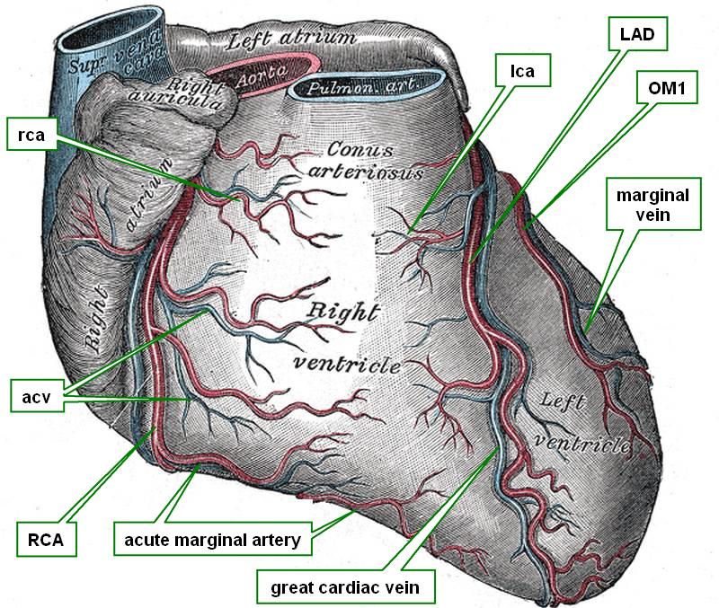
Anterior view of the heart
The great cardiac vein is one of the longest veins of the coronary system of the heart. It arises close to the cardiac apex in the anterior interventricular sulcus and ascends parallel to the anterior interventricular artery (LAD). The great cardiac vein will end in the most proximal portion of the coronary sinus. At this point the coronary sinus presents with an “entrance” valve called the “coronary valve of Vieussens”
In some cases the great cardiac vein will anastomose with the middle cardiac vein creating a collateral circulation venous drainage system that includes the coronary sinus.
There are several anatomical variations of the great cardiac vein.
Sources:
1 "Tratado de Anatomia Humana" Testut et Latarjet 8 Ed. 1931 Salvat Editores, Spain
2. “The great cardiac vein” Peikovic, B. Surg Radiol Anat. 1992;14(1):23-8.
3. “Great cardiac vein variations” Bales, GS Clin Anat. 2004 Jul;17(5):436-43p>Source:
4. "Anatomy of the Human Body" Henry Gray 1918. Philadelphia: Lea & Febiger
Image modified by CAA, Inc. Original image courtesy of bartleby.com
- Details
Medical suffix originating from the old Greek [πλήσσειν] (plessein) meaning “to strike” or the modern Greek [πληγείσες] meaning “stricken”. Its meaning in modern medical terminology is “paralysis”, the loss or impairment of bodily movement. This suffix can be seen in many medical terms:
- Quadriplegia: From the term [quad] meaning “four”. Loss of movement or paralysis in all four extremities
- Hemiplegia: From the prefix [hemi-] meaning “half”. Loss of movement or paralysis in one half of the body
- Paraplegia: The prefix [para-] means “alongside” or “parallel to”. Originally the word paraplegia was synonymous with [hemiplegia], but in modern times it is used to denote loss of movement or paralysis in the lower half of the body
- Cardioplegia: The root term [card] means “heart”. Stopping or paralyzing the heart. This is done as part of a heart operation where a solution is placed into the heart to stop it and allow for heart surgery. In the case of minimally invasive surgery retrograde cardioplegia is used by placing the cardioplegia solution through a catheter into the coronary sinus.
Note: The links to Google Translate include an icon that will allow you to hear the Greek or Latin pronunciation of the word.
- Details
Click for a larger image
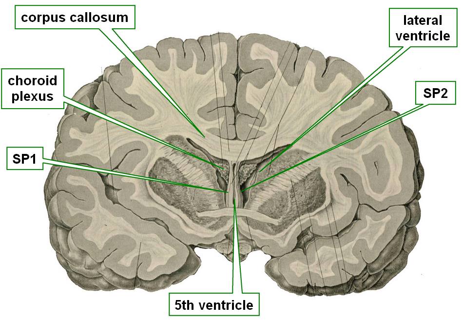
The septum pellucidum is a thin, transparent membrane that forms the medial wall of the lateral ventricles.
The term [septum] means “wall” or “partition” and [pellucidum] means “transparent”.
The septum pellucidum is found most of the time as a single, median structure that is shared by both lateral ventricles and is sometimes perforated, which has no pathological consequence.
In some cases this structure can be double, creating a separate medial wall for each lateral ventricle. When this happens the space between the lateral ventricles has been named the “cavum septi pellucidi”, the “fifth ventricle”, or is eponymically called the “ventricle of Vieussens”. This is shown in the accompanying image
Image modified from the original from “Surgical Anatomy” by John Deaver 1901. Public domain.
- Details
Click for a larger image
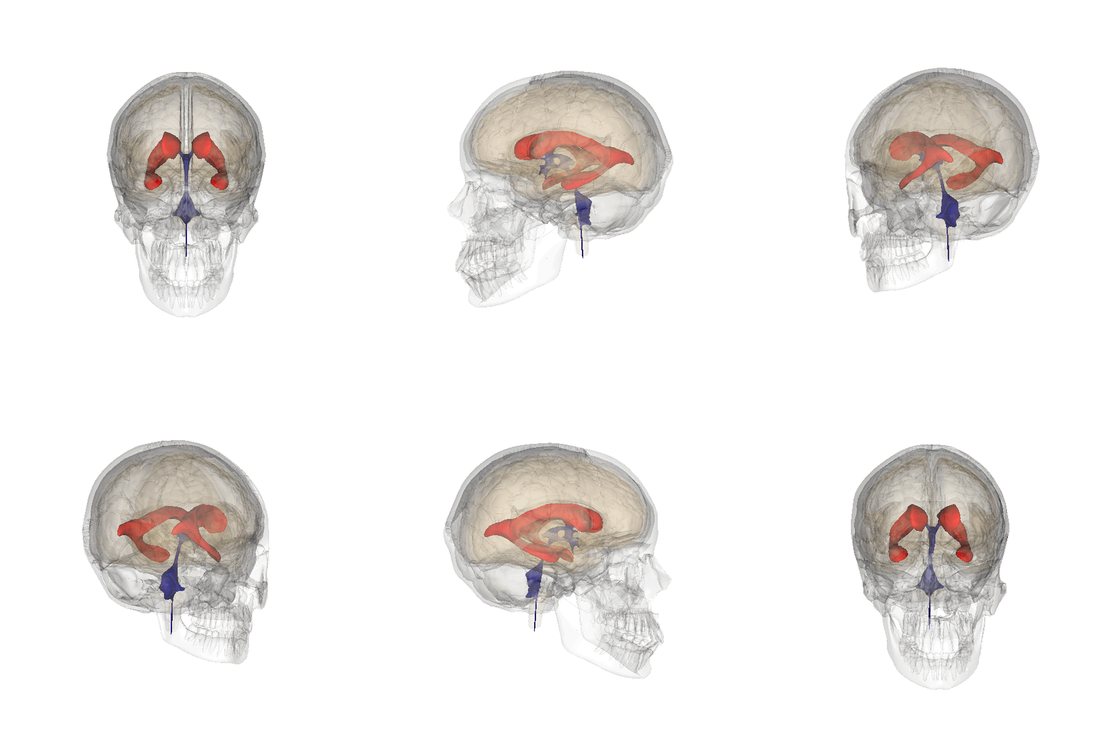
The lateral ventricles of the brain are two separate cavities, each found within a cerebral hemisphere. The lateral ventricles are part of the ventricular system of the brain and contain cerebrospinal fluid which is produced within the ventricular system in the choroid plexuses, most of which are found in the lateral ventricles.
Each lateral ventricle communicates with the third ventricle by way of an interventricular foramen, also known as the foramen of Monro, allowing for circulation of the cerebrospinal fluid
Each lateral ventricle has four components:
1. The central component or “body”. This area is found superior to the thalamus and inferior to the corpus callosum
2. An anterior extension called the “frontal horn”. The boundary between the body and the frontal horn is the interventricular foramen of Monro
3. A posterior extension called the “occipital horn”
4. An anteroinferior extension called the “temporal horn”
The body of the lateral ventricle and the occipital and temporal horn meet at a common point called the “ventricular crossroad” or “isthmus”.
The accompanying animated image shows the lateral ventricles in red. Click on the image for a larger depiction
Sources:
1 "Tratado de Anatomia Humana" Testut et Latarjet 8 Ed. 1931 Salvat Editores, Spain
2. "Gray's Anatomy" 38th British Ed. Churchill Livingstone 1995.
3. Image credits: By Images are generated by Life Science Databases (LSDB). (License information))], public domain, via Wikimedia Commons
- Details
The medical prefix [hemi-] means “half”. It is also the basis for the prefix [semi-] , also meaning “half”. We can see this term used in many medical terms such as:
- Hemisphere: literally “half a sphere”, as in the case of the cerebral hemispheres
- Hemiplegic: Paralized only on one half of the body
- Hemialgia: Pain on one half (of the body)
- Hemicephalalgia: Pain on one half of the head, a migraine
- Semilunar: Half a moon, name used for the aortic and pulmonary valves
- Details
Click for a larger image
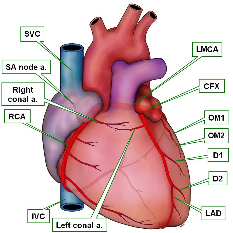
The eponym “ring of Vieussens” refers to a collateral circulation anastomotic communication between the right conal artery and the left conal artery. This communication, when present, is a potential life-saving pathway when there is stenosis or obstruction at the origin of either the right or the left coronary arteries, allowing blood to bypass the blockage.
This anastomosis is sometimes evident, although sometimes when the anastomosis is not seen on the surface of the heart, there is the possibility that the anastomosis is present subepicardially as demonstrated in the 2014 study by Loukas et al.
This ring is demonstrated in the accompanying image. For a three-dimensional volume–rendered CT demonstrating Vieussens’ collateral pathway please click here. For the full 2006 article by Hansen, click here.
Image property of CAA, Inc. Artist: Victoria Ratcliffe.